This awareness week for eye care is to encourage more people to have regular sight tests and make lifestyle choices that benefit their vision and general wellbeing.
Please go for an eye test.
There is so much more than opticians just giving you glasses to read or for distance.
They can detect eye problems early that could be prevented or treated. More serious conditions can also be discovered by the advancement and availability of the latest scanning or retinal photography in some branches.
In the past you could only be offered these things in hospital eye clinics or centres.
Your eyesight will be slightly different in each eye at the very least, so seeing an optician will help correct any problems individually with a made to measure prescription.
My own condition was seen at just a normal check up by an Asda optician. Who then referred me to the Eye Centre for further investigation.
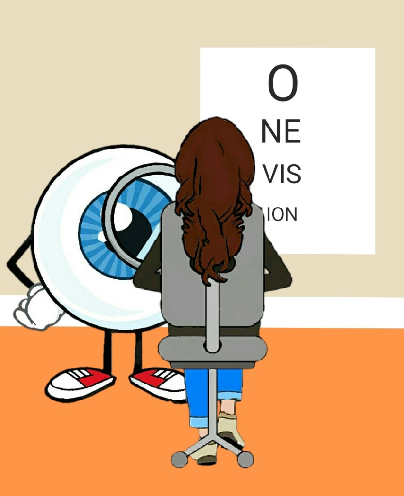
PICTURE DESCRIPTION Cartoon depiction of myself looking at an eye test on the wall. With One Vision written on it.
This is an epic blog post. Goes on abit. There’s a lot to tell you, but it Isn’t a rant. (Definitely a cuppa, and maybe a sandwich post)…
My urgent ish next hospital appointment was on Saturday 17th September 2022. 16 days after my meeting with doctor who requested these further tests.
I’ve surely had all the tests. I’ve been in most rooms, in 3 different hospitals, trying to find out the answers to something, sometimes, I don’t want the answers to.
I’ve had flashing tests, tests in the dark, reading tests, colorblind books, sat in different waiting rooms, numerous doctors shining lights through prisms for ages. wires, (yes… wires) drops, on and in my eyes.
One thing though. My eye phobia isn’t half as bad as it once was.
This time someone came with me. He’s visually impaired himself and uses a white cane. I just thought it would be funny to bring him along, which he kindly offered, so everyone would expect him to move when a names been called out to go for a test. They would think ‘oh bugger, it’s catching’, when they saw me get up instead, someone who hasn’t any obvious clues that I’m actually, to the people the waiting room, in THEIR gang.
We spoke about this and had a chuckle.
I absolutely love breaking peoples conceptions and stereotypes.
If we made some eyebrows raise, and a nudge to their mate. Then that’s all good.
He can see the time on his ‘very normal, not at all adapted’ watch.
I can’t.
See what I mean?
Don’t judge people.
Do you know that only 2 to 8% of the people who use a white cane, can see nothing at all?
See? You don’t just get ranty, sometimes long winded, sometimes funny (there will be more at somepoint… It’s gotten abit heavy recently) posts, occasionally there’s abit of sight loss facts too.
Anyway (I’m rambling again)…
I’m more positive this time and have an ‘is what, it is mentality’
I have the usual eye test. I’m still reading the same lines for both eyes. Any lower and I’m guessing, due to fuzziness, blurriness and missing tiny ‘bits’ Any of the above, a few things together, or all three.
“Am I seeing someone today?” I ask?
“No”…
I explain to the woman the whole boring never ending saga of me, waiting on results and waiting to see the main man.
She looks at the screen and sees that I’ve had the tests a while ago and yes, I’m waiting to see the big fella.
Oh believe me lady… I know this fact. She pops a note on the file.
I’m proper on a mission today and not at all being despondent, like I was at the previous appointment.
Then I’m straight into the Visual Field test which is a machine you look into, resting your chin on a blue stripe chin rest (left hand side) for your right eye to look for different sized white lights that flash intermittently, around what is, your peripheral vision.
You have to pop a pirate patch on your opposing eye.
You are given a clicky button. To click when you can see any light flashing on the screen.
If this was a game on the ZX Spectrum in the 80s, it would have been fab.
Takes a good 7 to 8 minutes for each eye to go through the process. When it’s your left eyes turn, you pop the patch onto your right eye and move your chin to a white stripe chin rest. On the right hand side.
It’s not uncomfortable at all. The lights aren’t that bright.
I did notice the difference between my clicking for the left and right eye.
I wasn’t as clicky fantastic, when my right eye was being tested.
Frustrating, as I don’t know if there were just less lights, or the fact there were the same amount and I just couldn’t see them.
Which, if you think about it, is the whole idea of the test.
PICTURE DESCRIPTION #1 Picture of a Visual Field test machine.PICTURE DESCRIPTION #2 Picture depiction of what you see using whilst having a Visual Field test. A grid with central red dot and single white lights appearing.
So after being a pirate for abit, I go find my buddy from the waiting room ‘cos we’ve got to go sit in the long, thin, unexpectedly empty, corridor. You know, the one with the Octopus Fields door in. Bloody Ringo. It’s now in my head… again.
There’s no dilation today. I think that’s because I’m not seeing the eye doc. My eyes won’t be examined today.
I go into a room next to the Octopus Fields door.
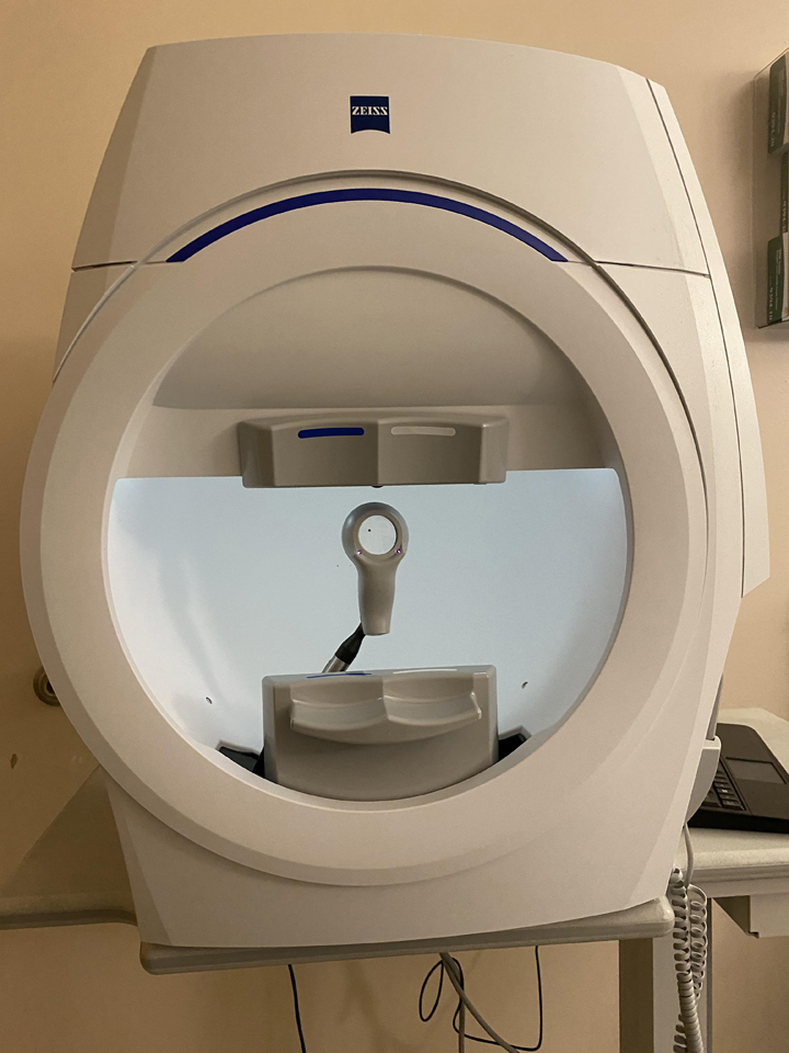
There’s another machine and this is for my OCT Disc scan.
PICTURE DESCRIPTION #3 Picture of an OCT Disc scan machine.
This machine has a red outline of a circle on a black background when you look into the eyepiece. Off to the side is a bright blue light in the shape of a cross that you have to stare at so the nurse can get a picture of your retinal disc.
PICTURE DESCRIPTION #4 Picture of what you see looking through the eyepiece on a OCT machine. Black background with red circle and blue cross in top left corner.
Then, very quickly, I’m done. Off for a pint or three.
Now waiting to see someone for these results of why my peripheral vision is dodgy. I’ve no idea when or who that will be with. At all.
My Consultant I’m waiting on, may have retired, just doesn’t want to see me or makes chutney for a living now for all I know anymore…
If you want to read about the OCT Disc scan. I’ve wrote a post about it, as I haven’t had one of these before. Please click link below.
I’ve had plenty of tests but only had this one on the 17th September, so thought I would explain about it incase anyones having a scan soon and wondering what to expect.
Optical Coherence Tomography (OCT) is a non-invasive imaging test. OCT uses light waves to take cross-section pictures of your retina.
With OCT, your eye doctor can see each of the retina’s distinctive layers. This allows your ophthalmologist to map and measure their thickness. These measurements help with diagnosis of several conditions.
What happens during OCT?
To prepare you for an OCT exam, your ophthalmologist may or may not put dilating eye drops in your eyes.
I didn’t have any put in myself.
These drops widen your pupil and make it easier to examine the retina.
You sit in front of the OCT machine and rest your head on a support to keep it motionless.
PICTURE DESCRIPTION #1 Picture of an OCT machine
You put each eye in turn, against the viewer to see the screen inside.
The equipment will then scan your eye without touching it. Scanning takes about 5 to 10 minutes.
The OCT scan uses light waves to create an image, similar to an ultrasound that uses sound waves to create an image.
The OCT will scan each of your eyes, whilst you fix your gaze on a blue cross in the top corner.
The red circle is for the retinal disc to fit into that the nurse will adjust from her side of things.
PICTURE DESCRIPTION #2 Picture of what you see looking through the eyepiece on a OCT machine. Black background with red circle and blue cross in top left corner.
OCT is often used to evaluate disorders of the Optic Nerve as well. The OCT exam helps your ophthalmologist see any changes to the fibers of the optic nerve.