The Guide Dogs for the Blinds new scheme – buddy dogs. Brings a new friend into the lives of children with sight loss. By helping to develop their self-confidence, improve relationships and build a greater sense of trust, these dogs can have a hugely positive effect on your child’s wellbeing – and your family, too.
These dogs weren’t suited to being a guide dog. This means you cannot use them as a guide dog or enter premises, shops with the dog.
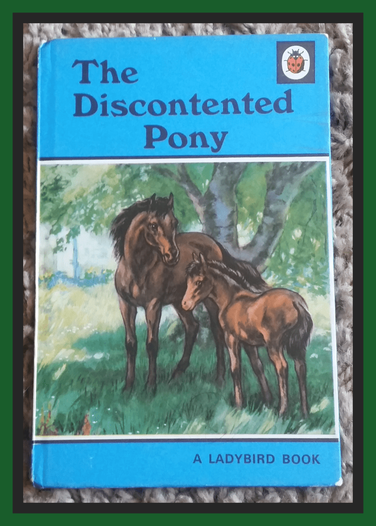
Thinking about my Mum today as it would have been her 81st birthday. I remembered my favourite book when I was a little girl.
I’ve got a collection of Ladybird Books. #geek. Ones that I remembered from when I was a child.
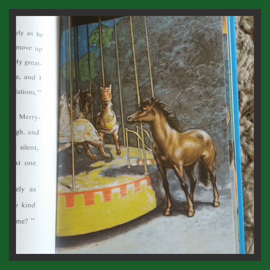
This is my favourite one. (Photo above. The Discontented Pony) I was so young though I couldn’t remember the story. The illustrations I remember vividly, as I suppose my Mum would have read it to me and I would have only looked at the pictures. I was obsessed with horses when I was a child. So loved this book.
I did used to like reading a proper book. Usually autobiographies, historical, crime or about space. Don’t really like fiction books at all. I would rather learn something real. I’ve still got some books I loved from my teenage years. Well worn but just didn’t have the heart to give them away.
So when it was too hard to read a book, book. I started to read them on my mobile phone. At least you can adjust the font. It became increasingly annoying though as you didn’t get many words to the page. You were just flicking it all the time.
Now I have a 10.1 inch screen tablet. This is loads better and I enjoy reading again for a little while until my eyes sort of say “Right, we’ve had enough now”
Either through pain developing around the sockets, a headache, or just everything becomes extremely blurry with double vision. All of a sudden. Just like that.
It’s cuppa tea time for this one I think… It’s long. Abit rambling… Get ready…
Masked up, I entered the eye centre with loads of time to spare. There are clear screens up around the reception. This is because of Covid.
Had to hold my letter up to show the woman behind the screen. Then went into the very sparsely chaired waiting room. It’s only small and usually packed with chairs. Today about 12 chairs were spaced out for social distancing.
They were having a book sale. Each book for 50p. Just normal print books. Instantly made me sad and reminded me that I can’t hold and read a proper book. I was bemused, did think to myself that it was taking the p** abit. It wasn’t probably the best place to put it. In the eye hospital… but then again, I wouldn’t have thought about that either, before this happened to me.
I do read on my big 10.1 tablet though. I’ve started reading about The Beatles at the moment. Just finished Elvis & Ginger.
Anyway… Went to have the usual eye test. The one you have at the opticians. Had to have pinhole glasses given to me for both eyes as I was guessing that the number 8 I saw, must be a letter S. These help the eyes focus abit more. I’ve done a post about pinhole glasses previously.
As far as I can remember. I think I was still reading on the same lines (3 and 4). So that’s good. Well. Not good. But you know what I mean.
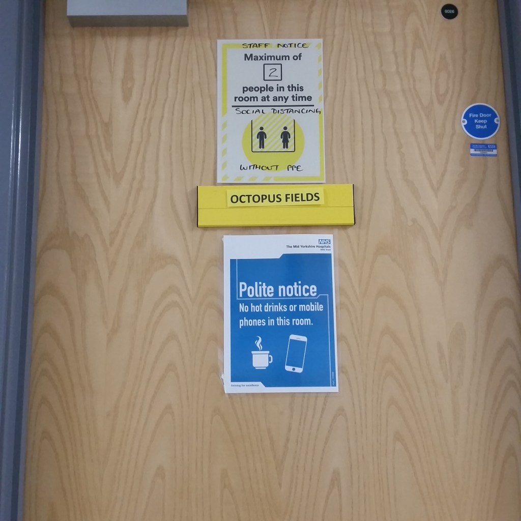
Then I went to sit in the sparsely chaired long corridor. Was told to sit infront of this door. (below). Cue. The tune Octopuses Garden, unfortunately entering my head.
Well. After being puzzled by what this appointment was for. I now knew I was off to the Octopus Fields.
Exciting ☺
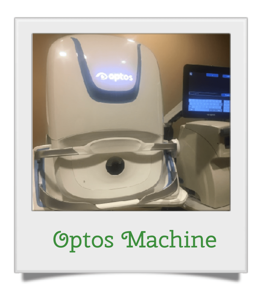
Name was called, so goes into the room. Was told to pop myself onto a wheely office chair in the middle of the room. #random. Then the bloke wheeled me the to the optos machine!
Weeeeeeeeee…
You place your right eye against the hole. Then colours appear. The green one gives out a massive flash. You can see for a split second your veins in your eye. Cue my outburst…
Me – “bloody hell”.
My Chauffer – (Laughing) “Oh, haven’t you had this test before?”
Me – “Clearly not”
Repeat with left eye.
All done very quickly. Didn’t have dilation drops for this one.
Then I was wheeled around again to another man to have another scan.
Again… Weeeeeeeeee.
This involved looking at a white dot while a red line lowers vertically, all while trying not to blink. I’m not sure what this is called. We were just chuckling at me being driven around on the chair for no reason whatsoever and I was trying my best not to blink. Didn’t think to ask.
So then that was it. Got my yellow sheet given to me. I asked if I was seeing the consultant. My chauffer with the chair for the day, explained that I wasn’t today and my other appointment I’m waiting for should come soon. Popped sheet into the tray at reception and off I went.
All in all. I think because of the zombie virus, and the fact we are still social distancing here in the UK. My appointment was quicker than usual, as there were less patients in the building. Within an hour I was done.
So… Ignorance is bliss. No news from this appointment. Will get a follow up letter though.
Now waiting on Glaucoma Field test. Then appointment to see the consultant.