Posted in My Tests Explained

What happens at a Visual field Test

A depiction of what you see in a field test. Square grid and dots
To check your field of vision.

I’ve had quite a few of these tests.

I’ve got all of the tiny lights so far.

You pop your face into the machine and then you focus on the central dot. Without moving your eyes from this position, you then tell the nurse how many little white dots you can see.

Posted in Follow up letters

Testing times

Pinhole glasses on a clipboard
I’ve found out what my tests are I’m having.
Got a follow up letter and appointment for this coming Tuesday today.
That was quick, and a bit unsettling if I’m honest.
I don’t really understand all the medical terms but, Right eye PED at macula (looked like a little hole dip in the layer) and both eyes show outer retinal atrophy.
The test I am having on Tuesday the 1st October is for Autofluorescence
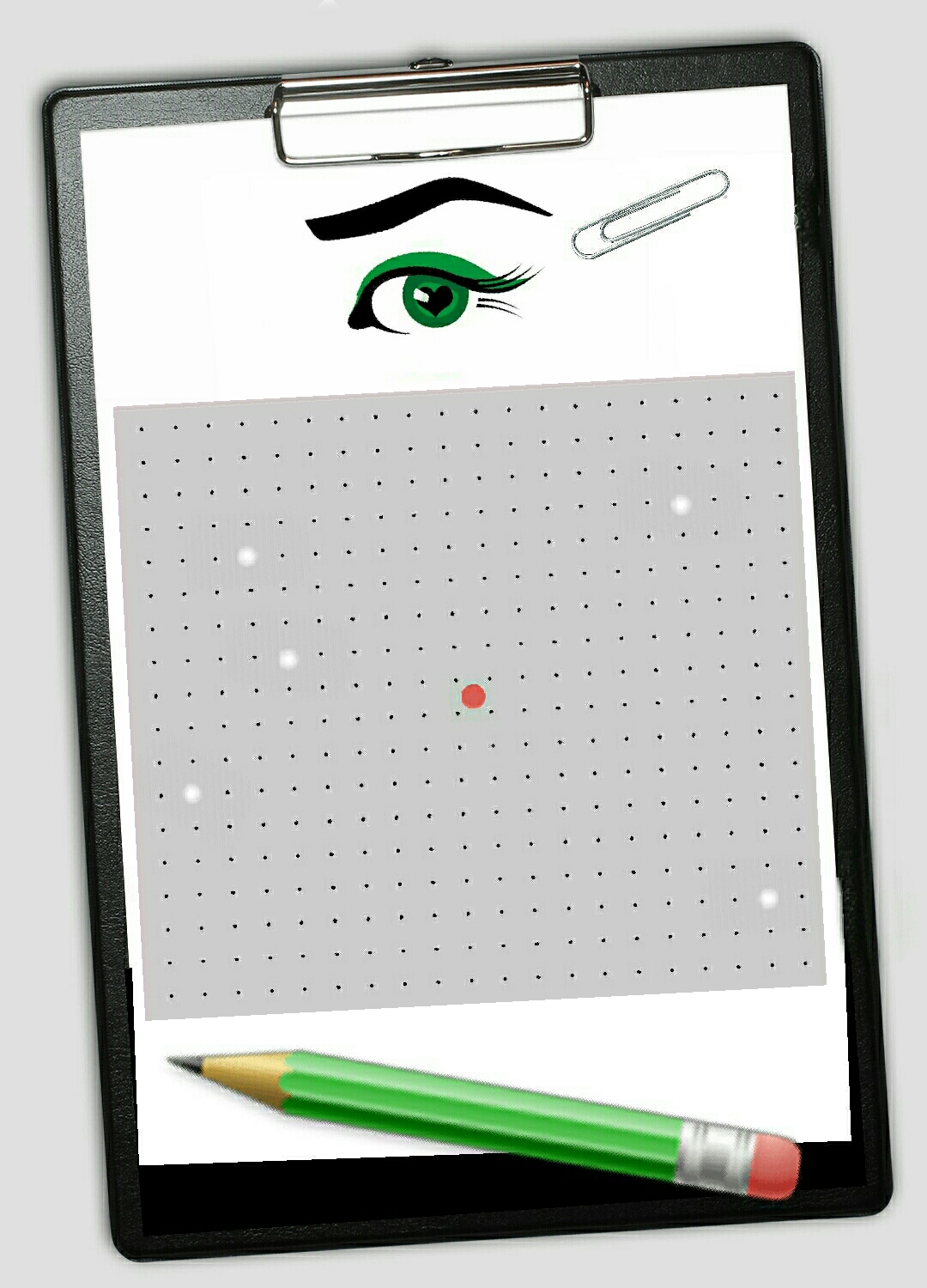
I also have one on the 3rd of October for a visual field test.
Then I’m waiting for an appointment for an ERG.
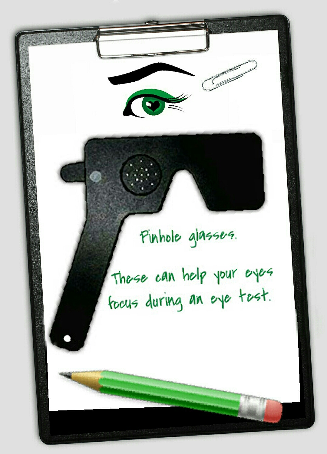
My right eye best corrected, (which means with distance glasses ) is 20/80. 20/60 with pinhole.
My left eye best corrected is 20/60. 20/50 with pinhole.
This is a change, from the 20/20 vision (corrected) when I was first diagnosed four and a half years ago. 20/30 2 years ago.
I feel worried and a bit sick if I’m honest.
It’s happening. This is really happening. Quicker than I thought it would.
Posted in My Tests Explained

What is an Autofluorescence Test?

Autofluorescence is the concept of using naturally occurring fluorescence from the retina to provide an overall picture, literally, of eye health.
Illuminating the retina with blue light causes certain cellular components to ‘glow’ without injecting any dye. This glow (fluorescence) returning from the retina can be used to create a black and white image which can be interpreted by the eye specialist by seeing any characteristic and common patterns in eye conditions.
Autofluorescence patterns may be linked to disease progression in patients with macular dystrophy and may thereby help consultants determine clear diagnosis and condition of eye health.
Use of this imaging is being used in a variety of retinal diseases including: age-related macular degeneration, retinitis pigmentosa and macular dystrophies.
I haven’t had this done yet. I have will be very interesting to see what’s in there.
Just looks like a black and white photograph. I won’t pop a photograph up. Looks a bit grim.